
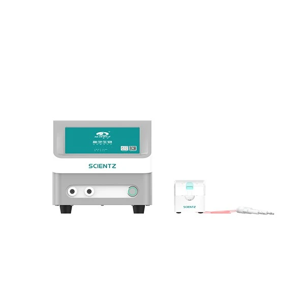
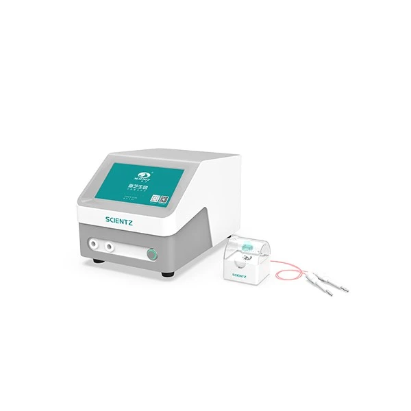
Electroporation, also known as cellular electroporation, is a key method for introducing exogenous macromolecules like DNA, RNA, siRNA, and proteins, as well as small molecules, into cells.
Under the influence of a transient strong electric field, the cell membrane becomes permeable, allowing charged exogenous substances to
enter the cell in a manner similar to electrophoresis. Due to the high resistance of the cell membrane’s phospholipid bilayer, the voltage across
the cell is mostly borne by the membrane, with minimal voltage in the cytoplasm. This leads to negligible cytotoxicity during electroporation
under normal conditions. Once DNA or other substances pass through the membrane, they stay near the membrane before the cell’s mecha
nisms transport them to the nucleus or other parts of the cell.
Since electroporation relies on physical methods, the molecular characteristics on the cell surface have minimal effect on the process. Unlike
chemical transfection or viral vector methods, electroporation can be applied to all cell types and is easily quantifiable.
Under the influence of a transient strong electric field, the cell membrane becomes permeable, allowing charged exogenous substances to
enter the cell in a manner similar to electrophoresis. Due to the high resistance of the cell membrane’s phospholipid bilayer, the voltage across
the cell is mostly borne by the membrane, with minimal voltage in the cytoplasm. This leads to negligible cytotoxicity during electroporation
under normal conditions. Once DNA or other substances pass through the membrane, they stay near the membrane before the cell’s mecha
nisms transport them to the nucleus or other parts of the cell.
Since electroporation relies on physical methods, the molecular characteristics on the cell surface have minimal effect on the process. Unlike
chemical transfection or viral vector methods, electroporation can be applied to all cell types and is easily quantifiable.
Cell Electroporation Electric Field Diagram
1 The cell membrane acts as an insulator, causing the electric current in the electroporation fluid
near the cell to distort.
2 In a series circuit, the larger the resistance, the higher the voltage; most of the voltage is borne by
the cell membrane.
3 Only one end of the cell is effectively electroporated.
4 The voltage in the cytoplasm is minimal; DNA halts at the membrane after electroporation and
slowly diffuses into the cell via cellular mechanisms.
5 The voltage at the nuclear membrane is extremely low, with no voltage inside the nucleus,
resulting in no genotoxicity during electroporation.


Technical Parameters
| Conversion Rates for Different Strains | ||||||||
| Type Vo | Pulse ltage(V) |
Resistance (Ω) |
Capacitance (μF) |
Electroporation Cup(mm) |
Pulse Type Voltage(V) |
Resistance (2) |
Capacitance (μF) |
Electroporation Cup(mm) |
| E.Coli | 1800 | 200 | 25 | 1 | S.pyogenes 2100 | 200 | 50 | 2 |
| E.Coli | 2500 | 200 | 25 | 2 | L.plantarum 2000 | 400 | 25 | 2 |
| E.Coli | 3000 | 200 | 25 | 2 | Eukaryotes | |||
| A.tumefaciens | 2400 | 200 | 25 | 1 | S.cerevisiae 1500 | 200 | 25 | 2 |
| P.asruginosa | 2500 | 200 | 25 | 2 | S.pombe 2300 | 200 | 25 | 2 |
| S.aureus | 2900 | 50 | 25 | 2 | C.albicans 1500 | 200 | 25 | 2 |
| B.cereus | 1000 | 200 | 25 | 2 | P.pastoris 2000 | 200 | 25 | 2 |